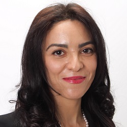
Faculty DirectoryLaleh Rad

Associate Professor of Biomedical Engineering and Radiology
Contact
2145 Sheridan RoadTech
Evanston, IL 60208-3109
Email Laleh Rad
Website
Departments
Electrical and Computer Engineering
Education
Post-Doctoral Fellow, Massachusetts General Hospital, Harvard Medical School, Boston, Massachusetts
Post-Doctoral Fellow, University of Toronto, Toronto, Canada
PhD in Electrical Engineering, Swiss Federal Institute of Technology (EPFL), Lausanne, Switzerland
MSc in Electrical Engineering, University of Tehran, Tehran, Iran
Research Interests
My lab focuses on application of computational modeling in medical imaging and medical device instrumentation. We are particularly interested in simulation-guided assessment and enhancement of safety of magnetic resonance imaging (MRI) in patients with implants, including but not limited to those with neuromodulation devices (such as deep brain stimulation systems) and cardiovascular electronic devices. We work hand in hand with major MRI vendors (Siemens) medical device companies (Abbott, Boston Scientific, Medtronic), FDA regulatory experts, and clinicians to devise novel surgical and technological methodologies to enhance safety of MRI in pediatric and adult patient populations with medical implants.
We are also interested in application of computational modeling and advanced neuroimaging to evaluate and guide neuromodulation therapies, with a focus on deep brain stimulation (DBS). Specifically, we create image-based patient-specific models of head and brain and use biophysical models of neural stimulation to quantify and optimize DBS therapeutic protocols.
Selected Publications
- Bhusal, Bhumi; Sanpitak, Pia Panravi; Jiang, Fuchang; Richardson, Jacob; Seiberlich, Nicole; Rosenow, Joshua M.; Elahi, Behzad; Golestanirad, Laleh, Comparative study of radiofrequency heating in deep brain stimulation devices during MRI at 1.5 T and 0.55 T, Magnetic resonance in medicine (2025).
- Yang, Huili; Aboyewa, Oluyemi B.; Webster, Gregory; Shah, Dhaivat; Golestanirad, Laleh; Baraboo, Justin J.; Markl, Michael; Collins, Jeremy D.; Knight, Bradley P.; Hong, Kyung Pyo; Patel, Amit R.; Lee, Daniel C.; Kim, Daniel, Aortic velocity measurements derived from phase-contrast MRI are influenced by a cardiac implantable electronic device in both adult and pediatric human subjects, Magnetic resonance in medicine (2025).
- Kutscha, Nicolas; Mahmutovic, Mirsad; Bhusal, Bhumi; Vu, Jasmine; Chemlali, Chaimaa; Hansen, Sam Luca J.D.; May, Markus W.; Knake, Susanne; Golestanirad, Laleh; Keil, Boris, A deep brain stimulation–conditioned RF coil for 3T MRI, Magnetic resonance in medicine (2025).
- Rad, Laleh Golestani, Adaptable MRI Coils for Enhanced Deep Brain Stimulation Imaging, Institute of Electrical and Electronics Engineers Inc. (2024).
- Zaidi, Tayeb; Marturano, Francesca; Bonmassar, Giorgio; Golestanirad, Laleh, Reduction of Medical Device Heating During Mri At 1.5 T and 3 T, IEEE Computer Society (2024).
- Tur, Yalcin; Vu, Jasmine; Waktola, Selam; Medetalibeyoglu, Alpay; Golestanirad, Laleh; Bagci, Ulas, Prediction of MRI-Induced Power Absorption in Patients with DBS Leads, Institute of Electrical and Electronics Engineers Inc. (2024).
- Henry, Kaylee R.; Ingersoll, Mark; Wartman, William; Jiang, Fuchang; Tang, Dexuan; Golestanirad, Laleh; Makaroff, Sergey N., An open-source user interface for real-time ultra-fast solving of electric fields around segmented deep brain stimulation electrodes, Brain Stimulation (2024).
- Vu, Jasmine; Bhusal, Bhumi; Rosenow, Joshua M.; Pilitsis, Julie; Golestanirad, Laleh, Effect of surgical modification of deep brain stimulation lead trajectories on radiofrequency heating during MRI at 3T, Journal of neurosurgery (2024).
Patents
Rad, L.G., Wald, L.L. and Bonmassar, G., General Hospital Corp, 2021. Mri-safe implantable leads with high-dielectric coating. U.S. Patent Application 16/970,550.
Rad, L.G., Bonmassar, G. and Pascual-Leone, A., General Hospital Corp, 2021. Systems and methods for ultra-focal transcranial magnetic stimulation. U.S. Patent Application 16/971,080.
Golestanirad, L. and Graham, S.J., Sunnybrook Research Institute, 2016. Electrode designs for efficient neural stimulation. U.S. Patent 9,526,890.