The Problem
Beam-sensitive zeolites are difficult to study at high resolution because traditional electron microscopy often damages or destroys their delicate crystal structures before meaningful data can be collected.
Beam-sensitive zeolites are difficult to study at high resolution because traditional electron microscopy often damages or destroys their delicate crystal structures before meaningful data can be collected.
Researchers applied near-axis transmission Kikuchi diffraction within a scanning electron microscope to map zeolite crystal orientations at the nanoscale while minimizing beam-induced damage.
The approach enables faster, safer, and more accessible characterization of zeolites, supporting advances in clean energy, environmental sustainability, and materials discovery.
PhD candidate Michael Barsoum; Professor Vinayak Dravid; Professor Omar Farha; Adjunct professor Steve Jacobsen; Tirzah Abbott, NUANCE Center SEM facility manager
Zeolites are not a household name, but they touch daily life in quiet and important ways. These porous crystals are used to capture carbon dioxide, refine fuels, soften water, and filter pollutants. Their effectiveness comes from an intricate internal structure full of nanoscale channels and cavities. Seeing that structure clearly is essential for improving how zeolites perform.
Before a recent Northwestern University study, that was not so easy.

New interdisciplinary research found a way to visualize fragile zeolite materials at the nanoscale without damaging them in the process. The work introduces an approach that allows scientists to safely and efficiently map crystal structures, opening new possibilities for clean energy, environmental sustainability, and materials discovery.
Zeolite crystals are typically synthesized in the lab through controlled chemical processes that form their characteristic porous frameworks. For this study, the crystals were prepared and supplied by the research team’s materials synthesis collaborators, allowing them to test the new imaging technique on well-known industrial zeolites.
“For decades, researchers have relied on powerful electron microscopes to examine materials at very small length scales. While these tools provide remarkable detail, they pose a problem for zeolites and other beam sensitive materials,” Professor Vinayak Dravid said. “The electron beam itself often damages or destroys the sample before a clear structural map can be collected. As a result, scientists have had to choose between resolution and preservation, limiting how much information could be gathered.”
The key issue is not whether the beam damages the crystal at all, but how much damage occurs before meaningful structural information can be obtained. Traditional transmission electron microscopy can deliver extremely high resolution, but in many cases, it degrades the zeolite before a complete dataset can be collected. This limits researchers’ ability to compare crystals, map defects, or link structure to performance.
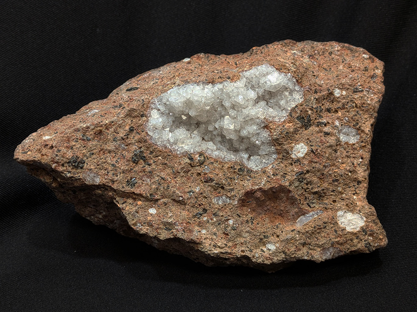
“Zeolites were first discovered in nature as secondary minerals formed during low-temperature alteration of mafic rocks like basalt,” said adjunct professor Steve Jacobsen, a mineralogist and co-author of the paper. “In lab-grown zeolites, we’re copying nature’s original design and adding specific functionality, but they’re just as susceptible to high-temperature degradation as natural ones because they have so much water in their structures.”
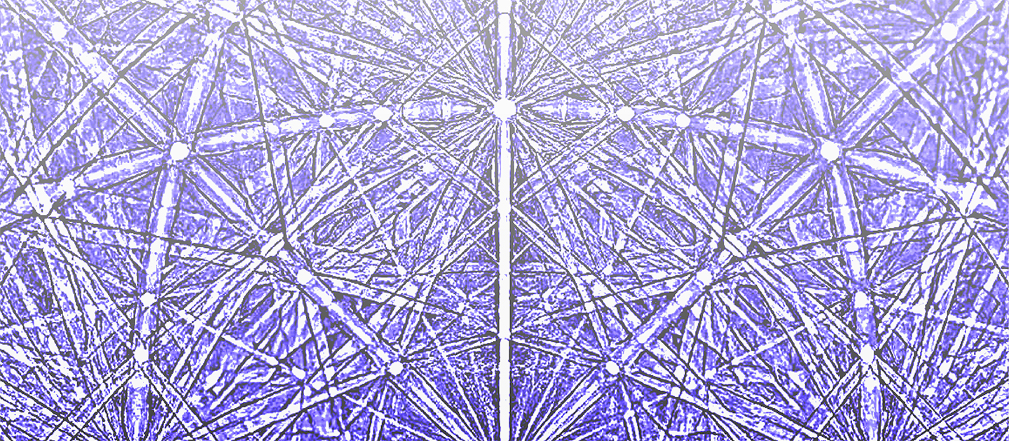
The Northwestern researchers addressed this challenge by applying a technique known as Near-Axis Transmission Kikuchi Diffraction (NA TKD) within a scanning electron microscope. Using the first detector of its kind in the United States, the researchers determined the crystal orientation of zeolite particles while minimizing beam damage. In practical terms, this means they could see how the crystals are arranged internally without compromising the material they were trying to understand.
The study focused on two widely used zeolites, ZSM-5 and Zeolite A. With NA TKD, the team achieved a spatial resolution of 75 nanometers, among the highest reported for this class of materials.
“This level of detail will allow researchers to link what a crystal looks like in a scanning electron microscope to its underlying structure,” Professor Omar Farha said. “It is an important step toward understanding why certain zeolites perform better than others in real world applications.”
The work was reported in the paper “Crystal Orientation and Defect Mapping in Electron-Beam-Sensitive Zeolites with Near-Axis Transmission Kikuchi Diffraction,” published earlier this month in the journal Nano Letters. Michael Barsoum, a joint PhD candidate in the labs of Dravid and Farha, was the paper’s first author. Jacobsen, an adjunct professor in the Weinberg College of Arts and Sciences’ Department of Earth, Environmental, and Planetary Sciences, was a co-author of the paper.
The paper was featured as a cover of the Feb. 18 edition of the journal.
Dravid is the Abraham Harris Professor of Materials Science and Engineering at the McCormick School of Engineering and (by courtesy) professor of chemistry and dermatology. Farha is Charles E. and Emma H. Morrison Professor in Chemistry and chair of the Department of Chemistry at Weinberg and (by courtesy) professor of chemical and biological engineering at McCormick.
Beyond resolution, speed is a key part of what makes the approach valuable. Traditional high-resolution techniques can be time consuming, sometimes allowing only a handful of particles to be analyzed in a session. In contrast, the NA TKD method is high throughput. Thousands of particles can be examined in minutes rather than hours. For the broader research community, this creates opportunities to combine microscopy with machine learning tools that rely on large datasets to identify patterns and guide the discovery of new materials.
The ability to rapidly and reliably map zeolite crystals has implications across multiple industries. Zeolites are foundational to the petroleum refining industry, where they catalyze key reactions. They are also critical in chemical manufacturing, water treatment, and air purification. In the clean-energy sector, zeolites are used in carbon capture and storage systems and as supports for catalytic processes in sustainable fuel production. By making structural analysis faster and more accessible, NA TKD could accelerate improvements in performance, lower production costs, and enable the design of new materials that are more efficient and durable.
The choice of a scanning electron microscope is also significant.
“Compared to transmission electron microscopes, SEMs are generally more cost effective and offer larger sample chambers,” Barsoum said. “This makes the technique more accessible and practical for routine characterization, particularly for beam sensitive zeolitic samples where phase purity and structural variation matter. “These rich datasets are also ripe for machine learning and AI pipelines for high throughput discovery and to push the limits of determining space group, possibly even structure, from raw patterns.”