Variety in Enamel Composition May Predict Later Tooth Health
Longer lifespans, disparities in dental health make understanding tooth enamel more important
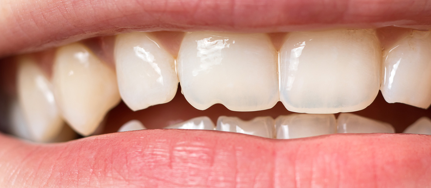
Tooth decay has forced unpleasant dental visits on most of the U.S. population. In fact, over 90 percent of adults have had at least one cavity. Furthermore, longer lifespans and egregious dental health disparities are contributing to rising cases.
Yet despite the work of biologists, materials scientists, chemists, engineers, and clinicians, not much is known about how or why some people are more likely than others to experience tooth enamel decay or congenital conditions.
Now, an interdisciplinary team of Northwestern University researchers has mapped ions within human teeth and found significant structural differences between samples that may enhance understanding of enamel’s life cycle and impact on human health.
A new paper published December 19 in the Proceedings of the National Academy of Sciences demonstrates new approaches to measuring differences in tooth composition at a previously unresearched, hard-to-measure scale of tooth enamel.
Tooth enamel may be one of the most complex materials produced by vertebrates — a significant barrier to more fully understanding teeth in the context of formation and overall well-being.
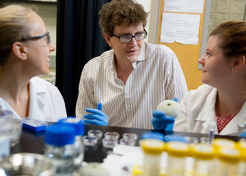
“This paper fits into the broader goal of understanding the structure of this important biological material and its role in human health, especially in light of dental cavities and tooth decay,” said Robert Free, the paper’s first author and a materials science and engineering researcher at Northwestern Engineering.
Using X-ray diffraction technology, the team was able to observe tiny differences — on the order of picometers, a fraction of the size of a single atom — in the crystallographic parameters of enamel crystallites, which they linked to the presence of ions like magnesium and sodium or carbonate.
“There are very few techniques that can measure these particular ions within enamel crystallites accurately,” said the McCormick School of Engineering’s Derk Joester, the paper’s corresponding author. “They have low atomic numbers and concentrations, so ultimately, we predict compositional differences that are on the order of a fraction of an atomic percent. This requires extreme accuracy and is only possible with X-rays using a lateral approach to predict composition.”
Joester is an associate professor of materials science and engineering at McCormick. He is a member of Northwestern’s Chemistry of Life Processes Institute and the Materials Research Center.
In the study, the researchers looked at three healthy teeth from human adults. Technology including synchrotron X-ray diffraction and unsupervised machine learning then revealed structures within the mesoscale features, specifically, single enamel rods and the surrounding inter-rod enamel.
Rods are bundles of around 10,000 crystallites — very thin, long crystals — that are stacked together in an orderly fashion like a fistful of dry spaghetti. Previously, working with atom probe tomography and transmission electron microscopy, researchers could only look at a small area within a rod without the ability to distinguish differences between crystalline lattice parameters in rod and inter-rod enamel.
This paper fits into the broader goal of understanding the structure of this important biological material and its role in human health, especially in light of dental cavities and tooth decay.
Robert FreeMaterials Science and Engineering Researcher, Northwestern Engineering
The team discovered differences in the lattice orientation and structure between enamel samples at the mesoscale level. The mesoscale is an intermediate scale that lies between the atomic scale and those observable by the human eye. Where other scales are well understood, without more advanced technology, the mesoscale cannot be measured. A longstanding partnership with the Advanced Photon Source (APS), a Department of Energy Office of Science user facility at Argonne National Laboratory, enabled the Northwestern team to investigate structures at the mesoscale, where rods can be captured.
“The APS is a huge resource to Northwestern,” said Stuart Stock, a research professor of cell and developmental biology at Northwestern University Feinberg School of Medicine and an author on the paper. “There are all these sophisticated machines at Argonne that give our students the opportunity to use cutting-edge tools frequently without having to jump on an airplane each time.”
Looking at different ions and seeing where they’re localized in the enamel tells us what cells are doing during formation and what they’re contributing.
Stock described the study's sample size as a "Goldilocks" population because it demonstrated new findings, taught the team how to prepare notoriously difficult enamel samples and showed them there's that this scale can reveal about things like tooth formation and general health.
“We’ve raised more questions than we started with,” Stock said. “We haven’t totally laid out and described it, but there’s something going on here, and there needs to be a lot more work done at this scale on this particular problem.”
The researchers have developed new theories on teeth based on key findings of the study. For example, rods looked different than anticipated and had significant variation between each individual's tooth. Whether the variations are common to all humans or a critical piece of evidence to why populations including women are more susceptible to tooth decay remains to be seen.
The team believes answers to these questions could lead us to an understanding of how tooth enamel develops, which would be critical knowledge for scientists developing enamel in labs or attempting to stop tooth decay before it begins.
“Seeing the inorganic fingerprint of enamel crystals gives us insight into what was happening when the enamel was formed,” said Victoria Cooley, a PhD student who studies animal models of teeth in Northwestern’s materials science department. “Looking at different ions and seeing where they’re localized in the enamel tells us what cells are doing during formation and what they’re contributing.”
Magnesium and carbonate ions, for example, control solubility of enamel, so their composition in tooth enamel may be critical to how and when teeth break down. With findings that there are differences in enamel structure attributed to changes in lattice parameters, the team thinks composition between teeth differs, too, marking a step closer to the heart of tooth decay.