Method to Control Cellular Structures Could Lead to Better Cancer Treatment
Lipids’ ability to split used to create spatially organized regions of protein presentation on nanoparticles
Architecture and design are important in all aspects of life. Whether it’s constructing a major building or creating new products, projects must be meticulously planned to succeed.
That’s especially true for therapies to treat cancer, even if it is incredibly difficult to control cellular structures at the nanoscale. But using simple methods, a team of researchers led by Northwestern Engineering’s Neha Kamat devised an approach that leverages lipids’ natural ability to separate to create spatially organized regions of protein presentation, which could make an underperforming cancer treatment more effective.
“This technique could help scientists develop better nanoparticle therapeutics, especially those that rely on interacting with cell membrane receptors because many cell receptors are sensitive to the spatial organization of the ligands and proteins they interact with,” Kamat said. “It's also interesting to think about how cells are nature's first engineers, and they need functional design just like we do.”
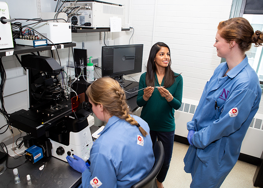
Kamat is an assistant professor of biomedical engineering at the McCormick School of Engineering. Timothy Vu, a graduate student in Kamat’s lab, was the first author of the paper “Lipid Phase Separation in Vesicles Enhances TRAIL-Mediated Cytotoxicity,” which was recently published in Nano Letters.
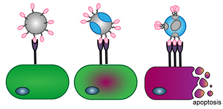
Using the natural ability of lipids — think olive oil and butter — to separate on the surface of a lipid nanoparticle, the investigators created those spatially organized regions of protein presentation that could enhance the effectiveness of TNF-related apoptosis-inducing ligand (TRAIL) treatment. TRAIL is a naturally occurring protein that immune cells express to kill cancer cells, but has only shown limited efficacy in clinical trials.
“TRAIL's limited efficacy might be because the TRAIL protein used as a therapeutic does not resemble TRAIL on a cell membrane,” Vu said. “We created nanoparticles (lipid vesicles) that mimic the lipid rafts in a cell membrane, which enhances TRAIL's ability to kill certain cancer cells.”
This work represents a step forward because, while phase-separated lipid vesicles have been studied for decades, their use in nanomedicine has been sparse. Kamat and her collaborators showed that phase-separated lipid vesicles, where lipids that have different physical properties phase separate into different regions on a vesicle membrane, can be used to enhance the amount of damage to cancer cells inflicted by a conjugated therapeutic protein like TRAIL. There’s also the possibility this type of nanoparticle presentation can enhance the potency of other therapeutic proteins like antibodies.
In the future, the research team will explore how the phase-separated lipid vesicles act in the body.
“It is known that the lipid composition of vesicles can affect what cells interact with the vesicles or what organs the vesicles will accumulate to, but we don't have any data for phase separated lipid vesicles,” Vu said. “It would be interesting if by mixing different lipids, we get completely different biodistribution. We are also extending this technology to create nanoparticle antibody-mimetics, and to see how phase-separated lipid vesicles could enhance therapeutic antibodies.”