Materials Scientists Drill Down to Vulnerabilities Involved in Human Tooth Decay
Enamel formation study could lead to new interventions to prevent and treat disease and defects
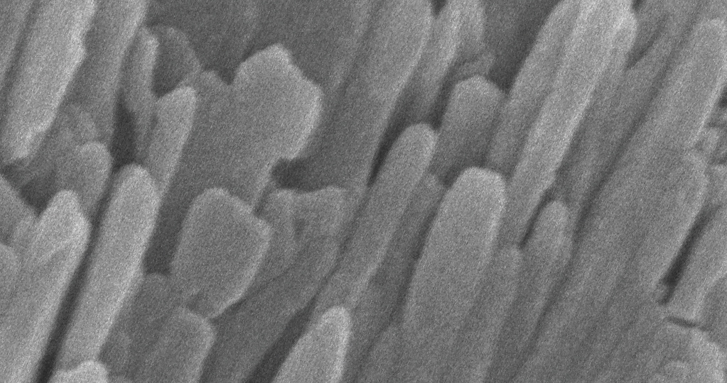 Enamel is made of tightly bunched, oblong crystals that have a width about 1,000 times smaller than a human hair.
Northwestern Engineering researchers have cracked one of the secrets of tooth decay. In a new study of human enamel, the materials scientists are the first to identify a small number of impurity atoms that may contribute to the enamel’s strength but also make the material more soluble. They also are the first to determine the spatial distribution of the impurities with atomic-scale resolution.
Dental caries -- better known as tooth decay -- is the breakdown of teeth due to bacteria. (“Caries” is Latin for “rottenness.”) It is one of the most common chronic diseases and a major public health problem, especially as the average life expectancy of humans increases.
 The McCormick School of Engineering discovery in the building blocks of enamel -- with detail down to the nanoscale -- could lead to a better understanding of human tooth decay as well as genetic conditions that affect enamel formation, which can lead to highly compromised or completely absent enamel.
Enamel, the human tooth’s protective outer layer, covers the entire crown. Its hardness comes from its high mineral content.
“Enamel has evolved to be hard and wear-resistant enough to withstand the forces associated with chewing for decades,” said Derk Joester, who led the research and is an associate professor of materials science and engineering. “However, enamel has very limited potential to regenerate. Our fundamental research helps us understand how enamel may form, which should aid in the development of new interventions and materials to prevent and treat caries. The knowledge also might help prevent or ameliorate the suffering of patients with congenital enamel defects.”
The study was published July 1 by the journal Nature. The title of the paper is “Chemical Gradients in Human Enamel Crystallites.”
Joester is the corresponding author. Karen A. DeRocher and Paul J.M. Smeets, a PhD student and a postdoctoral fellow, respectively, in Joester’s lab, are co-first authors. In addition to Joester, DeRocher and Smeets, other authors of the paper are Linus Stegbauer, Michael J. Cohen, Lyle M. Gordon, and McCormick School of Engineering associate professor of materials science and engineering James M. Rondinelli, of Northwestern; Berit H. Goodge, Michael J. Zachman and Lena F. Kourkoutis, of Cornell University; and Prasanna V. Balachandran, of the University of Virginia.
One major obstacle hindering enamel research is its complex structure, with features across multiple length scales. Enamel, which can reach a thickness of several millimeters, is a three-dimensional weave of rods. Each rod, approximately five microns wide, is made up of thousands of individual hydroxylapatite crystallites that are very long and thin. The width of a crystallite is on the order of tens of nanometers. These nanoscale crystallites are the fundamental building blocks of enamel.
Perhaps unique to human enamel, the center of the crystallite seems to be more soluble, Joester said, and his team wanted to understand why. The researchers set out to test if the composition of minor enamel constituents varies in single crystallites.
Using cutting-edge quantitative atomic-scale techniques, the team discovered that human enamel crystallites have a core-shell structure. Each crystallite has a continuous crystal structure with calcium, phosphate and hydroxyl ions arranged periodically (the shell). However, at the crystallite’s center, a greater number of these ions is replaced with magnesium, sodium, carbonate and fluoride (the core). Within the core, two magnesium-rich layers flank a mix of sodium, fluoride and carbonate ions.
“Surprisingly, the magnesium ions form two layers on either side of the core, like the world’s tiniest sandwich, just six-billionths of a meter across,” DeRocher said.
Detecting and visualizing the sandwich structure required scanning transmission electron microscopy at cryogenic temperatures (cryo-STEM) and atom probe tomography (APT). Cryo-STEM analysis revealed the regular arrangement of atoms in the crystals. APT allowed the researchers to determine the chemical nature and position of small numbers of impurity atoms with sub-nanometer resolution.
The researchers found strong evidence that the core-shell architecture and resulting residual stresses impact the dissolution behavior of human enamel crystallites while also providing a plausible avenue for extrinsic toughening of enamel.
“The ability to visualize chemical gradients down to the nanoscale enhances our understanding of how enamel may form and could lead to new methods to improve the health of enamel,” Smeets said.
This study builds on an earlier work, published in 2015, in which the researchers discovered that crystallites are glued together by an extremely thin amorphous film that differs in composition from the crystallites.