

Engineering Technology Empowers Engagement
Technologies and methods developed by Northwestern Engineering research teams are helping widen worlds.


Imagine waking up in the morning seeing sunshine stream through your window and hearing the coffee machine gurgling in the kitchen. You stretch, get up out of bed, and walk down the stairs. After a quick breakfast, you open your front door and walk outside, ready to face another day.
It’s a routine that many people take for granted. But when one of these abilities—sight, hearing, touch, mobility—is taken away, a simple routine can become challenging, possibly insurmountable.
That’s where engineers can make a difference. At Northwestern, researchers are developing new technologies and methods to prevent blindness, restore hearing, facilitate feeling through touch, improve the ability of wheelchair users to better navigate their environments, and help stroke survivors walk.
The goal? To empower people with the tools they need to engage successfully with their surroundings and one another.

Optimizing the Human Health Span
The technologies featured in this article restore the senses and mobility that make independent living possible, advancing our priority of Optimizing the Human Health Span. At Northwestern Engineering, we seek not only to extend how long people live, but how fully they can engage with the world around them.
Sight
Goal: Prevent blindness due to glaucoma and help make whole-eye transplants viable
Researchers

Hao F. Zhang
Professor of Biomedical Engineering
Director of the Center for Engineering in Vision and Ophthalmology
Nicholas J. Volpe
George W. and Edwina S. Tarry Professor of Ophthalmology
Cheng Sun
Professor of Mechanical Engineering
Mark Johnson
Professor of Biomedical Engineering
Glaucoma, a common eye disease and a leading cause of blindness in older adults, develops when high fluid pressure in the eye damages the optic nerve. To facilitate better drainage, surgeons typically implant stents in the inner nasal angle in the front of the eye, the most accessible location. Unfortunately, that location does not always provide the best drainage. As a result, high pressure often recurs.
At the Center for Engineering in Vision and Ophthalmology (CEVO), Professor Hao Zhang and his collaborators are committed to translating engineering discoveries into ophthalmology practice. They are exploring how to combine imaging with AI to model the individual patient’s unique form to find optimal stent locations guided by the eye’s many fluid outlets, called collector channels.

“Each eye is different,” Zhang says. “We should implant stents at locations based on the features of each eye.”
To develop such a model, Zhang and his team use noninvasive imaging that shines light within the front of the eye to build a 3D replica of its internal anatomy. They then use physics-guided AI tools to build a numerical model that can predict the outcomes of placing one or more stents in different locations.
With support from the Forsythe Family Foundation, Zhang and his collaborators are using human donor eyeballs and animal models to train the software to build the AI models. Their goal is to demonstrate to clinicians how this approach benefits patients.
“Our collaborating physicians tell us that this is a longstanding challenge, and our hope is to have the data to guide surgical planning within four or five years,” Zhang says.
Zhang’s team isn’t content to stop there. For glaucoma patients who have become blind from retinal neurodegeneration, no current treatment exists to restore sight.
Zhang and his collaborators from across the United States are working on a federally funded project to make whole eye transplants a reality. To do so, researchers must figure out how to regenerate the functional optic nerve that connects the eye to the brain. Zhang’s innovative imaging technologies are being used to determine how long an eyeball can remain viable after it is removed from the body and whether neurons have actually regenerated after controlled damage.
“It’s a moonshot project,” Zhang says. “Nobody knows whether it’s going to be successful or not, but during the process, there will be many new tools developed, which will help patients regardless.”
The potential for new discoveries in this area could one day benefit the treatment of other ailments. “With the right imaging data, with the right AI agent, clinical management and diagnosis of the eye will be able to impact the whole health of the human body, including heart, brain, and more,” Zhang says.
Hearing
Goal: Develop cochlear implants that offer better hearing than current devices
Researchers

Petia Vlahovska
Professor of Engineering Sciences and Applied Mathematics
Claus-Peter Richter
Professor of Otolaryngology at the Feinberg School of Medicine
Professor of Biomedical Engineering (by courtesy)
Much of human communication, learning, and awareness happens through hearing. For people who are deaf or severely hard of hearing, cochlear implants can relay a sense of sound to the brain by stimulating the auditory nerve.

Such implants, however, are imperfect. By electrically stimulating neurons with metal electrode arrays, they create sounds that can seem off and nonspecific. Because researchers assumed neurons fire only through electrical stimulation, these implants have remained the only option.
If the membrane itself changes state during stimulation, then that’s a mechanism we can engineer around to build better hybrid cochlear implants for people who have run out of options with today’s devices. Professor of Otolaryngology at the Feinberg School of Medicine, Professor of Biomedical Engineering
Professor Petia Vlahovska knows that a different sort of implant is possible. At Northwestern University’s Feinberg School of Medicine, Professor Claus-Peter Richter has shown that action potentials—tiny sparks that neurons use to send signals throughout the body—can also be stimulated with infrared light instead of electricity. Other research has shown that action potentials are not only electrical but also mechanical waves.
For Vlahovska, an applied mathematician, the idea that neurons could be stimulated differently has opened a whole new way of thinking about machine-brain interfaces. Her previous research into the behavior of soft materials has led her to study cellular membranes, which she now hopes to harness to create new types of implants.
“If we don’t need electricity to propagate signals, could we do it mechanically through soft materials that better connect with tissue?” she asks.
With a $3 million Trailblazer Engineering Impact Award from the National Science Foundation, she and Richter aim to do just that. Using Vlahovska’s expertise in fluid mechanics, membranes, and biophysics and Richter’s expertise in developing cochlear implants, the team will create and test synthetic membrane systems that generate the dynamics of action potentials without using electrical currents.

Together, they will study whether the infrared stimulation works by heating nerve membranes to trigger stimulation. Confirming that could lay the groundwork for future neuroprosthetic technologies, including better cochlear implants.
“I’ve been circling this question since my master’s thesis, and it’s exciting to finally have the collaborators and the resources to answer it directly,” Richter says. “If the membrane itself changes state during stimulation, then that’s a mechanism we can engineer around to build better hybrid cochlear implants for people who have run out of options with today’s devices.”
Touch
Goal: Improve foot sensing for people with peripheral neuropathy
Researcher

John Rogers
Louis Simpson and Kimberly Querrey Professor of Materials Science and Engineering, Biomedical Engineering and Neurological Surgery
Sensations on the bottoms of human feet give people information about their environment, movements, and even balance. When nerves are damaged—whether from diabetes or stroke—diminished foot sensation can lead to more trips and falls.
Wearable sensor expert John Rogers wondered if he could combine the flexible sensor technology he’s developed with miniaturized forms of haptic technology, which use tactile responses to give feedback to users. Such a system could help people with this sort of peripheral neuropathy sense the ground beneath their feet in a new way.
“For people who are unable to sense the pressure on their feet during normal walking and movement, we thought that we might be able to reproduce those patterns of pressure as tactile sensations on another part of the body, as a form of sensory substitution,” Rogers says. “If we could build a soft, wearable technology to serve that purpose, then users might be able to effectively learn how to feel their feet through their upper legs or forearms, thereby enabling them to walk with more confidence and without risks of falling.”

Working with researchers at the Shirley Ryan AbilityLab and the Georgia Institute of Technology, he and his team developed a shoe insole outfitted with an array of sensors that quantifies the pressure distribution on the foot in real time as users walk. That pattern of pressure is then wirelessly transmitted to actuators placed on a person’s forearms or upper thighs.
Each actuator gives off a small vibration, which corresponds with pressure from different parts of the foot. That essentially allows users to feel on their forearms or upper thighs what they would normally feel on the bottom of their foot. When the device was tested on stroke survivors at the AbilityLab, users could translate the vibrations accurately within an hour or two.

“With this type of system, you don’t need to look at your feet while you’re walking,” Rogers says. “You are free to walk normally, looking at your surroundings and perhaps having a conversation with a friend. Tests with patients showed that even a modest amount of training with these devices led to significant improvements in their gait and balance.”
This sensory substitution technology goes beyond feet. Working with a veteran who lost his lower arm in combat, Rogers’s team developed a system that included a robotic hand with an array of pressure sensors embedded into the fingers. Those sensors transmitted the information to haptic arrays mounted on his upper arm, giving him a sense of “touch” that allowed him to pick up and manipulate objects by “feeling” them on his upper arm. The device was so successful that he could pick up an eggshell without cracking it.
Next, the team will work to simplify the insole technology using fewer sensors and actuators to reduce the cost and complexity of the system without sacrificing its efficacy. They are also exploring other potential uses. “Adding this haptic capability across the wide range of sensors that we’ve already developed could profoundly increase the therapeutic value to patients,” Rogers says.
Mobility
Goal: Help wheelchair users drive with independence
Researcher

Brenna Argall
Professor of Mechanical Engineering, Computer Science, and Physical Medicine and Rehabilitation
Principal Investigator of Project DRIVE
Wheelchair users steer through their environments using a variety of controls—including joysticks, head movements, and sip-and-puff switches. Yet each of these controls can only be integrated into most wheelchairs through one kind of proprietary cable.
Software and communication protocols developed by rehabilitation robotics expert Brenna Argall and her collaborators in Northwestern’s Project DRIVE have made it easier to integrate any digital interface with wheelchairs. Project DRIVE focuses on wheelchair driving autonomy, accessibility, and independent mobility, and has produced the first-ever open-source wheelchair digital interface (WDI).
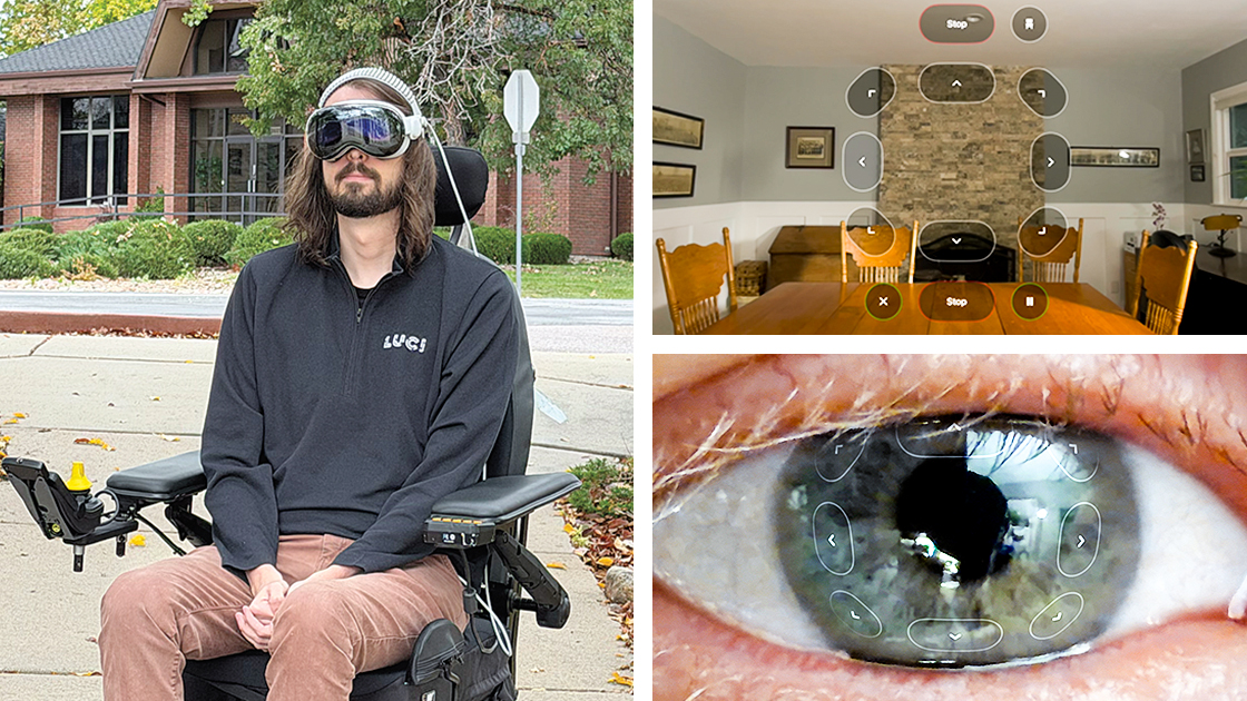
With our WDI, you can use an Xbox controller to drive your wheelchair or even a USB clicker to indicate an emergency stop—any technology with a USB or Bluetooth interface,” Argall says.
In September, Apple released a new accessibility feature leveraging the precise eye-tracking of Apple Vision Pro, empowering users of compatible alternate drive systems to control powered wheelchairs via the WDI using only their eyes.
“Apple Vision Pro users have the option to control a wheelchair with just their eyes, demonstrating how technology can expand independence for people with disabilities,” Argall says. “The wheelchair digital interface is open source and accessible to anyone, so hopefully this will help the wheelchair industry adopt this as a standard.”
The WDI was developed collaboratively by Project DRIVE with partners LUCI, a physical AI company that brings smart technology to powered wheelchairs, and LifeDrive Mobility, an alternative controls partner.
Supported with funding from the National Science Foundation, the Project DRIVE team hasn’t stopped there: Argall’s lab has developed an algorithm that will help users of wheelchairs equipped with LUCI’s technology better avoid obstacles. Currently, these wheelchairs slow or stop when approaching an obstacle. The new algorithm would help the wheelchairs actively drive around obstacles. The researchers also are working to develop fully autonomous wheelchair driving on top of LUCI’s deterministic safety layer.
“Most wheelchair users want to stay in control, but some users, such as those with late-stage ALS, want full autonomy in certain contexts,” Argall says. “We keep pushing, because we know there’s more we can do for wheelchair driving. We want people to have driving systems that meet their needs.”

Goal: Redefine how stroke survivors relearn to walk
Researchers

Kevin Lynch
Professor of Mechanical Engineering
Director of Northwestern’s Center for Robotics and Biosystems
Jose Pons
Scientific Chair at Shirley Ryan AbilityLab
Professor of Physical Medicine and Rehabilitation at the Feinberg School of Medicine
Professor of Mechanical Engineering (by courtesy)
Daniel Ludvig
Research Scientist at Shirley Ryan AbilityLab
Research Assistant Professor at the McCormick School of Engineering
Levi Hargrove
Scientific Chair of the Regenstein Foundation Center for Bionic Medicine at Shirley Ryan AbilityLab
Professor of Physical Medicine and Rehabilitation at the Feinberg School of Medicine
Professor of Biomedical Engineering (by courtesy)
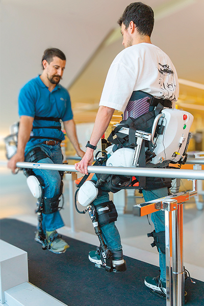
When stroke survivors relearn to walk, they often practice on a treadmill with their weight supported by a harness. A physical therapist helps them lift and move their affected legs.
In recent years, robotic lower-body exoskeletons that give users extra strength have emerged as a possible rehab tool. Robotics expert Kevin Lynch and his collaborators at the Shirley Ryan AbilityLab recognized that putting patients into exoskeletons alone wouldn’t suffice—they would still need a physical therapist to guide them. But what if the therapist could guide them through their own separate exoskeleton?
Addressing that question, the team developed a new system called therapist-exoskeleton-patient interaction (TEPI), where both the patient and the therapist wear lower-body exoskeletons that are connected by virtual springs and dampers at the hips and knees. This enables the therapist to guide the patient’s movements.
“By moving their own legs to help move the patient’s legs, therapists have direct influence on the patient’s joint motions, as opposed to trying to control joint motions by using their hands to push or pull a particular location on the leg,” Lynch says. “It’s more intuitive for the therapist and also safer for the therapist long term, because they’re not bent over in an unnatural position, manually moving the patient’s legs.”
Therapists can also calibrate the strength of the virtual connection, which creates more personalized rehabilitation experiences for each patient. “That personalization allows the therapist to apply their own judgment and expertise while leveraging state-of-the-art robotics,” Lynch says.
By moving their own legs to help move the patient’s legs, therapists have direct influence on the patient’s joint motions, as opposed to trying to control joint motions by using their hands to push or pull a particular location on the leg. Professor of Mechanical Engineering, Director of the Center for Robotics and Biosystems
The system was tested on stroke survivors at the AbilityLab. With TEPI, patients had higher muscle activation and typically reached step heights and lengths 30 to 50 percent greater than they had using conventional manual therapy. Patients also reported high levels of satisfaction with the therapy.
The next step is for Northwestern and the AbilityLab to validate the system with many patients. The goal is to give therapists another tool to help patients recover.
“This sort of innovation is possible with Northwestern’s history of collaborative robot research and our close connection to the AbilityLab,” Lynch says. “It’s a big part of our mission and heritage.”